


Wide field retinal photographs are useful in both diagnosing and monitoring retinal conditions, and is used in conjunction with the Dr Chauhan’s examination on the microscope. Whilst an examination on a microscope allows for a 103 degree static view of the retina, the colour photograph allows a 200 degree static view. This technology is far superior to previous photography methods that only allowed for a maximum 45 degree view.
Wide field retinal photographs are performed in a similar method to traditional retinal photography, where your eye is placed in front of a camera lens and a bright flash is used to illuminate your retina during the imaging process. High quality images are able to be captured by our specialised cameras without the aid of pupil dilating drops.
Traditional Retinal Photography - 45 Degree
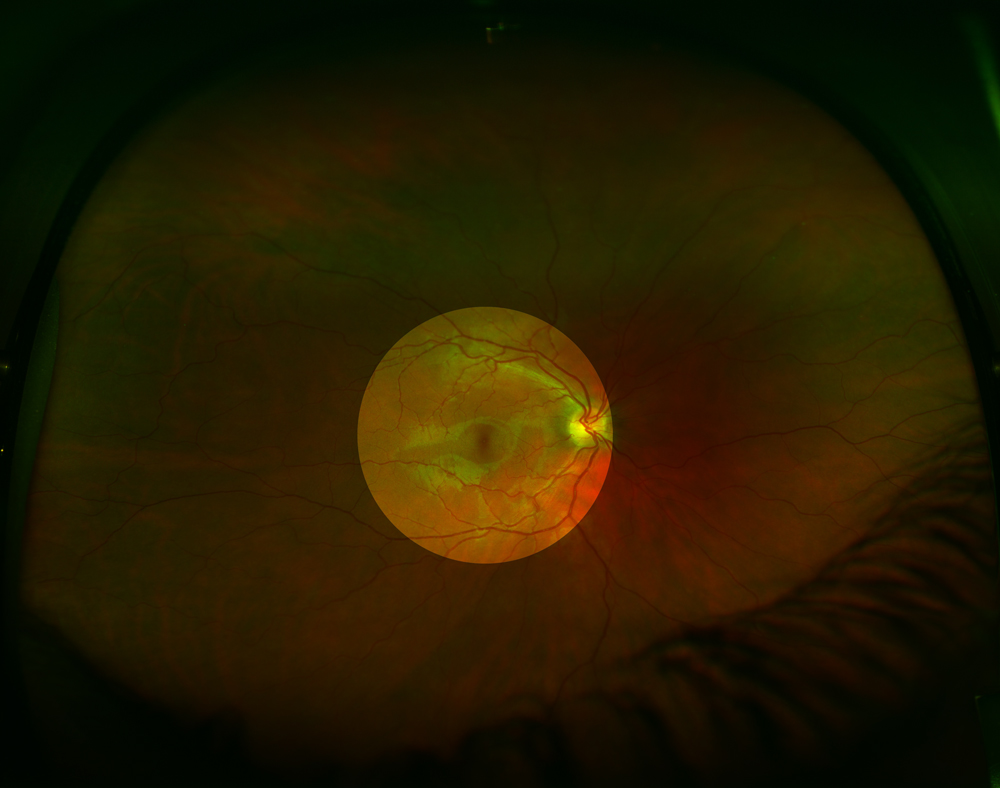 Traditional retinal photography provides only a 45 degree view on the retina (as highlighted in the centre of the picture). More peripheral areas of the retina can be photographed, although always only showing 45 degrees at a time (similar to shining a torch around in a dark room).
Wide Field Retinal Photography
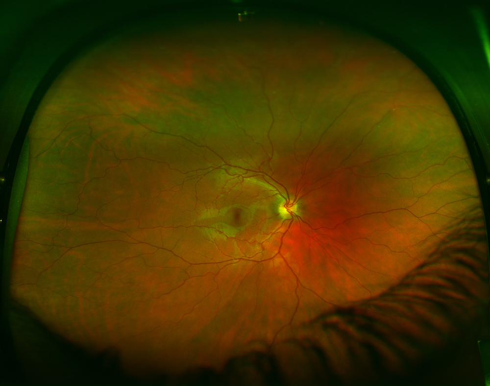 Wide field retinal photography provides an uninterrupted 200 degree view of the retina, documenting both the central and far peripheral areas of the retina.
The view is so wide that artefacts (such as eyelashes) can be caught in the photo – these are seen in the bottom right hand corner of the photo.