


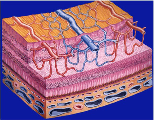
Cross-section of the retina and underlying choroid showing the different layers of blood vessels (courtesy of Novartis)
About Retinal Blood Vessels
The retina has two sources of oxygen and nutrients: the retinal blood vessels and the choroid, which lies under the retinal pigment epithelium.
The blood vessels within the retina itself that carry oxygen and nutrients are called arteries. The main one, the central retinal artery, enters the eye through the optic nerve and splits into the superior (upper) and inferior (lower) branches. These then keep branching out more, like the branches of a tree, until they form a very fine network of very thin blood vessels called capillaries.
It is mainly at the capillaries that oxygen and nutrients leave the blood, entering the retina, and that carbon dioxide and waste products leave the retina and pass into the blood to be taken away. Most of the problems caused by conditions affecting retinal blood vessels do so by either blocking these capillaries or causing them to become leaky. The capillaries join up to form branch veins and these then join at the optic nerve to form the central retinal vein that dives into the optic nerve on its way towards the heart.
Importantly, any part of the retina is only supplied by one artery and drained by one vein. As a result, if there is blockage of a retinal vein or artery, only the area of retina, and so only that part of the visual field, served by that blood vessel is affected.